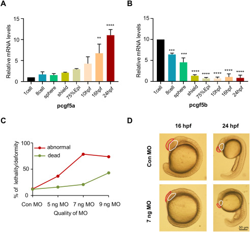
Fig. 6 Knockdown of pcgf5a expression results in abnormal early embryonic development in zebrafish. (A-C) qPCR and Real-time PCR was used to detect the expression of pcgf5a and pcgf5b in zebrafish embryos at different periods, from the 1-cell stage to 24 hpf. (D) A dose-dependent statistical graph of the embryo phenotype, and 7 ng MO was selected as the appropriate injection dose. (E) The phenotypes of zebrafish embryos taken under light microscope at 16 hpf and 24 hpf stage. Red markers show that the brain size of the pcgf5a-deficient zebrafish embryos is obviously smaller, and white markers show that the eye vesicles of the pcgf5a-deficient zebrafish embryos are smaller and the eye morphology is abnormally developed. Scale bar, 50 ?m.
Image
Figure Caption
Figure Data
Acknowledgments
This image is the copyrighted work of the attributed author or publisher, and
ZFIN has permission only to display this image to its users.
Additional permissions should be obtained from the applicable author or publisher of the image.
Full text @ Heliyon

