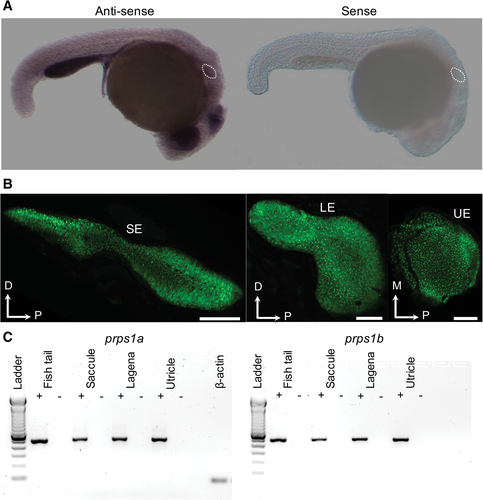
Fig. 2 (A) The prps1a expression in the otic vesicle of zebrafish. In situ hybridization whole mounts of 1-dpf zebrafish (wild-type AB) with the prps1a antisense (left) and sense (right) mRNA probes. With the prps1a antisense probe, specific moderate expression was observed in the entire embryo body, with more intense signals seen in the eyes, brain, gut, and otic vesicles, compared with the sense probe stain. The otic vesicles of the embryos are marked by dots. (B) Saccular epithelium (SE), lagenar epithelium (LE), and utricular epithelium (UE) of otolith organs of Et(krt4:GFP) sqet4 adult zebrafish. Green dots in the epithelia are hair cell bodies expressing GFP. D, dorsal; M, medial; and P, posterior. Scale bar = 100 ?m. (C) DNA gels of RT-PCR products from saccular, utricular, and lagenar epithelia of the Et(krt4:GFP) sqet4 adult zebrafish, showing the prps1a and prps1b expression in all three sensory epithelia of otolith organs. +: with reverse transcriptase, and ?: without reverse transcriptase. ß-actin was used as a positive control.
Image
Figure Caption
Figure Data
Acknowledgments
This image is the copyrighted work of the attributed author or publisher, and
ZFIN has permission only to display this image to its users.
Additional permissions should be obtained from the applicable author or publisher of the image.
Full text @ Anat. Rec. (Hoboken)

