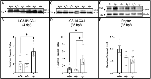
Fig. 9 Loss of raptor increases autophagy by 36 hpf, when there are reduced levels of Raptor protein. (A-D) Immunoblot analysis of Lc3 protein at (A,B) 4 dpf and (C,D) 36 hpf. (B,D) Graphs showing the ratio of Lc3-II/Lc3-I relative to wild type. (E,F) Immunoblot analysis of total Raptor protein at 36 hpf in wild types (circles), heterozygotes (triangles) and mutants (squares). Gapdh was used as loading control. Protein level was plotted relative to wild type. The statistical analyses were conducted using one-way ANOVA followed by Tukey's honest significant difference (HSD) post-hoc test for multiple comparisons, *P<0.05. Data are mean±s.e.m.
Image
Figure Caption
Figure Data
Acknowledgments
This image is the copyrighted work of the attributed author or publisher, and
ZFIN has permission only to display this image to its users.
Additional permissions should be obtained from the applicable author or publisher of the image.
Full text @ Development

