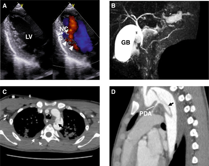
Echocardiography and magnetic resonance cholangio-pancreatography (MCRP) of the patient with the ACTA2 G148R variant. A An echocardiogram showing the left ventricular long-axis view during diastole. The ventricular wall of the left ventricle exhibits an outer compacted layer (C) and an inner non-compacted trabecular layer (NC), with deep recesses formed by the trabeculations (NC/C ratio 2.0). Color Doppler imaging shows blood infiltrating into the spaces between the trabeculae (arrowheads). B On MRCP, the main pancreatic duct exhibits a reverse Z-shape (arrows) in the head of the pancreas region. C, D Chest magnetic resonance imaging shows the descending aortic deviation. LV left ventricle, C compacted layer, NC non-compacted layer, GB gallbladder, PDA patency of the ductus arteriosus
Acknowledgments
This image is the copyrighted work of the attributed author or publisher, and
ZFIN has permission only to display this image to its users.
Additional permissions should be obtained from the applicable author or publisher of the image.
Full text @ J. Hum. Genet.
Your Input Welcome
Thank you for submitting comments. Your input has been emailed to ZFIN curators who may contact you if
additional information is required.
Oops. Something went wrong. Please try again later.