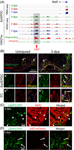
Fig. 6 LEN directs injury-induced Epi/EPDC expression. (A) Genome browser tracks of the genomic region near lepb showing the transcripts and chromatin accessibility profiles in the Epi/EPDC. The whole-ventricle H3K27Ac profile of the uninjured and regenerating heart is shown at the bottom. Gray box and red arrow indicate LEN. (B-D) Immunostained section images of transgenic fish carrying LENP2:EGFP. (B) Raldh2 antibody is used to label EC and Epi/EPDC. Uninjured heart shows one single Raldh2+ cell layer outlining the cardiac chamber. Raldh2 signal emerges in the ECs at the wound area and EPDCs in the cortical layers upon injury, which are co-labelled with LENP2:EGFP (Arrows). The boxed areas are enlarged at the bottom panels. (C) Myosin heavy chain (MHC) antibody is used to label CMs. (D) tcf21:mCherry is used for Epi/EPDC expression. While LENP2:EGFP rarely colocalizes with MHC+ CMs (Arrowheads), a subset of LENP2:EGFP co-localizes with tcf21:mCherry (Arrows). Note that asterix indicates CM expression as a basal expression of the P2 minimal promoter. At least five hearts for uninjured and injured samples were examined and all animals displayed a similar expression pattern
Image
Figure Caption
Acknowledgments
This image is the copyrighted work of the attributed author or publisher, and
ZFIN has permission only to display this image to its users.
Additional permissions should be obtained from the applicable author or publisher of the image.
Full text @ Dev. Dyn.

