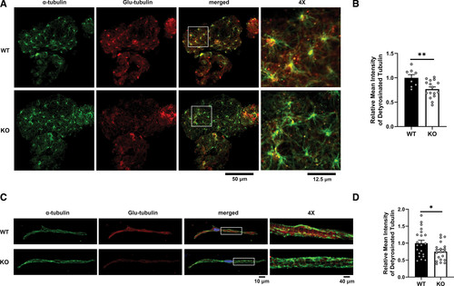
Fig. 5 mapre2 loss of function leads to decreased detyrosinated tubulin. A, Representative immunostaining of hearts from wild-type (WT; n=8) and homozygous knockout (KO; n=16) larvae showing a decrease in ventricular detyrosinated tubulin (Glu-tubulin) relative to total ?-tubulin. B, Quantification of ventricular Glu-tubulin signal using ?-tubulin signal as a mask (unpaired t test, P=0.0072). C, Representative immunostaining of ventricular myocytes isolated from adult WT (21 cells from 2 fish) and homozygous KO fish (20 cells from 3 fish) also showing a decrease in ventricular detyrosinated tubulin (Glu-tubulin) relative to total ?-tubulin. D, Quantification of ventricular Glu-tubulin signal using ?-tubulin signal as a mask (unpaired t test, P=0.0232). Representative images were chosen based on closeness to group mean and image quality.
Image
Figure Caption
Acknowledgments
This image is the copyrighted work of the attributed author or publisher, and
ZFIN has permission only to display this image to its users.
Additional permissions should be obtained from the applicable author or publisher of the image.
Full text @ Circ. Res.

