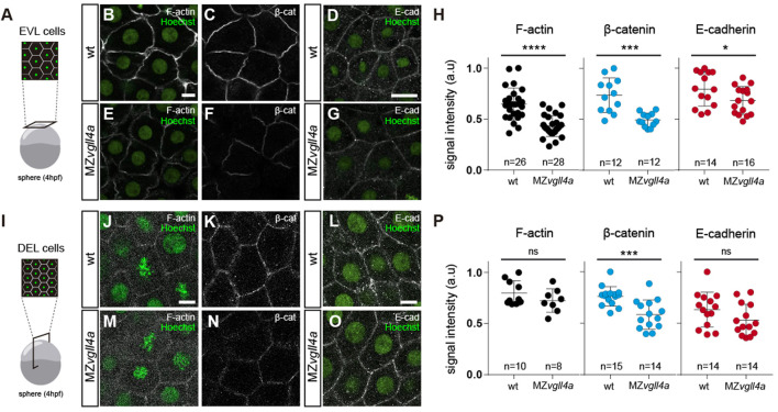
Maternal vgll4a is required for plasma membrane localization of the E-cadherin/β-catenin complex. (A,I) Schematic representation of the different imaging strategies. (B-G; J-O) Confocal images of F-actin (B, E, J, M), β-catenin (C, F, K, N) and E-cadherin (D, G, L, O) distribution in EVL (B–G) and DEL (J–O) cells in wt and MZvgll4a embryos at sphere stage. Embryos were counterstained with Hoechst (nuclei, green). (H,P) The graphs depict the fluorescent signal intensity (in arbitrary units, a. u) for F-actin, β-catenin and E-cadherin in EVL (H) or DEL (P) cells of wt and MZvgll4a embryos. Mann Whitney test. (H) β-catenin, p = 0.0001, E-cadherin; p = 0.0425. (P) F-actin, p = 0.3154; β-catenin, p = 0.0006 and E-cadherin, p = 0.1599. Ns, not significant, *p < 0.05; ***p < 0.001; ****p < 0.0001. Scale bar, 10 µm.
Acknowledgments
This image is the copyrighted work of the attributed author or publisher, and
ZFIN has permission only to display this image to its users.
Additional permissions should be obtained from the applicable author or publisher of the image.
Full text @ Front Cell Dev Biol
Your Input Welcome
Thank you for submitting comments. Your input has been emailed to ZFIN curators who may contact you if
additional information is required.
Oops. Something went wrong. Please try again later.