IMAGE

Fig. 1

ID
ZDB-IMAGE-240304-8
Source
Figures for Stegmann et al., 2024
Image
Figure Caption

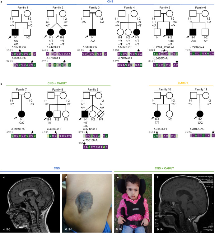
Fig. 1 Families with bi-allelic variants in CELSR3 and clinical images.

a Pedigrees of six families (1–6) with a predominant central nervous system (CNS) phenotype. b Pedigrees of three families (7–9) with combined CNS phenotype and congenital anomalies of the kidneys and urinary tract (CAKUT), and two families (10, 11) with CAKUT only. The evolutionary conservation of the affected sequence (bp) was estimated with the ConSurf server from variable (green) to conserved (purple). Asterisks: Position of the respective variants. The arrows indicate probands. Filled shapes should reflect affected status. c Brain magnetic resonance image (MRI) of 4: II-3 showing pachygyria. d Photograph of 6: II-1 showing a congenital hairy melanocytic nevus with a diameter of 0.1 to 0.15 meter at the level of the lower lumbal spine. Radiologic imaging of the spine was not performed here. e Photograph of 8: II-1 showing macrocephaly, high and prominent forehead and very small and low-set ears. f MRI of 9: II-1. Arrow: Chiari malformation type 1 (cerebellar tonsillar herniation).

Acknowledgments
This image is the copyrighted work of the attributed author or publisher, and ZFIN has permission only to display this image to its users. Additional permissions should be obtained from the applicable author or publisher of the image. Full text @ NPJ Genom Med