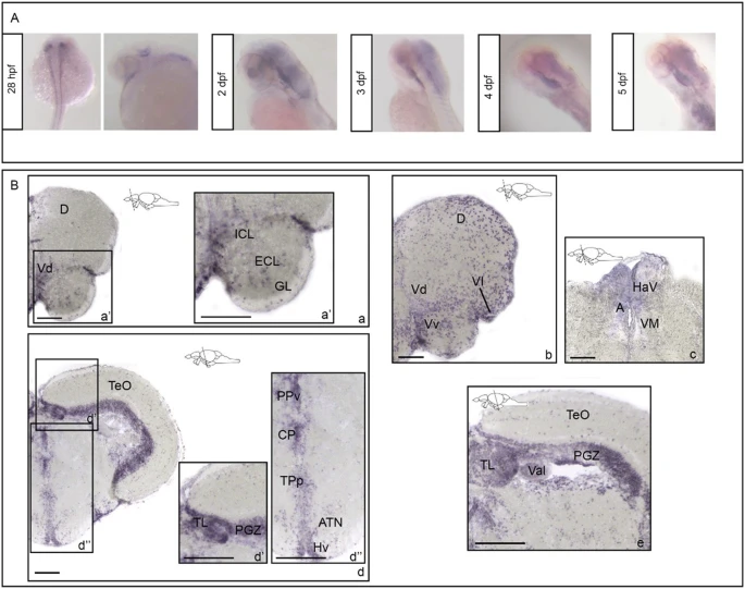
Fig. 1 rbfox1 shows restricted neuronal expression during development and is localised to specific forebrain, midbrain and hindbrain areas during adulthood.rbfox1 in situ hybridisation on (A) zebrafish whole mount larvae and (B) adult zebrafish brains, TL background. A rbfox1 whole mount in situ hybridisation on zebrafish larvae at 28 h post fertilisation, 3-, 4- and 5-days post fertilisation. B rbfox1 in situ hybridisation on adult zebrafish brains, (a?c) forebrain and (d?e) midbrain transverse sections. A anterior thalamic nuclei; ATN, anterior tuberal nucleus; CP, central posterior thalamic nucleus; D, dorsal telencephalic area; GL, glomerular cellular layer; HaV, ventral habenular nucleus; Hv, ventral zone of periventricular hypothalamus; ICL, internal cellular layer; PGZ, periventricular grey zone; PPv, ventral part of the periventricular pretectal nucleus; TeO, optic tectum; TL, torus longitudinalis; TPp, periventricular nucleus of posterior tuberculum; Val, valvula cerebelli; Vd, dorsal nucleus of ventral telencephalic area; Vl, lateral nucleus of ventral telencephalic area; VM, ventromedial thalamic nuclei; Vv, ventral nucleus of ventral telencephalic area. Scale bars: 100 µm (a, b, c, d); 200 µm (a?, d?, d?, e).
Image
Figure Caption
Figure Data
Acknowledgments
This image is the copyrighted work of the attributed author or publisher, and
ZFIN has permission only to display this image to its users.
Additional permissions should be obtained from the applicable author or publisher of the image.
Full text @ Transl Psychiatry

