
Image
|
Figure Caption
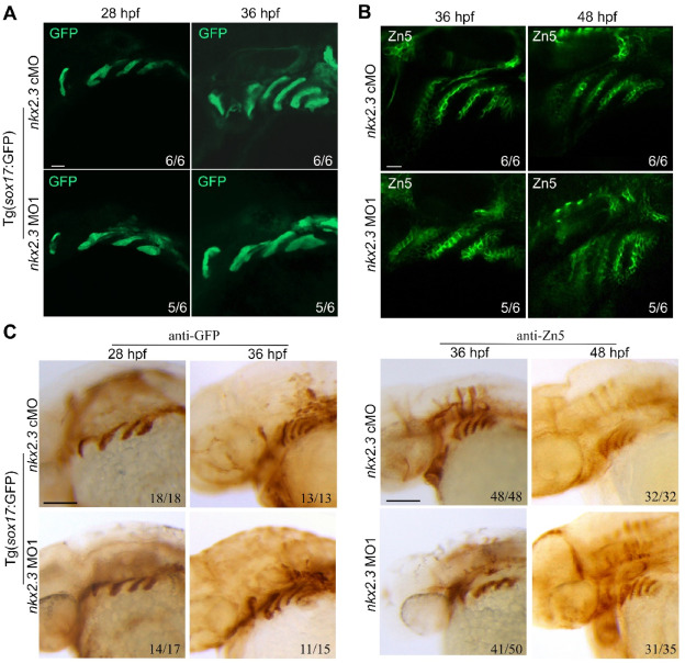
Fig. 3 Pharyngeal pouches are normally developed in nkx2.3 morphants. (A) Embryos were immunostained with the anti-GFP antibody at the indicated stages. Scale bars, 20 ?m. (B) Embryos were immunostained with the anti-Zn5 antibody at the indicated stages. Scale bars, 20 ?m. (C) Embryos were immunostained with the anti-Zn5 antibody at the indicated stages. Scale bars, 100 ?m. Images are shown with a lateral view and with the anterior to the left.
Figure Data
Acknowledgments
This image is the copyrighted work of the attributed author or publisher, and
ZFIN has permission only to display this image to its users.
Additional permissions should be obtained from the applicable author or publisher of the image.
Full text @ Heliyon