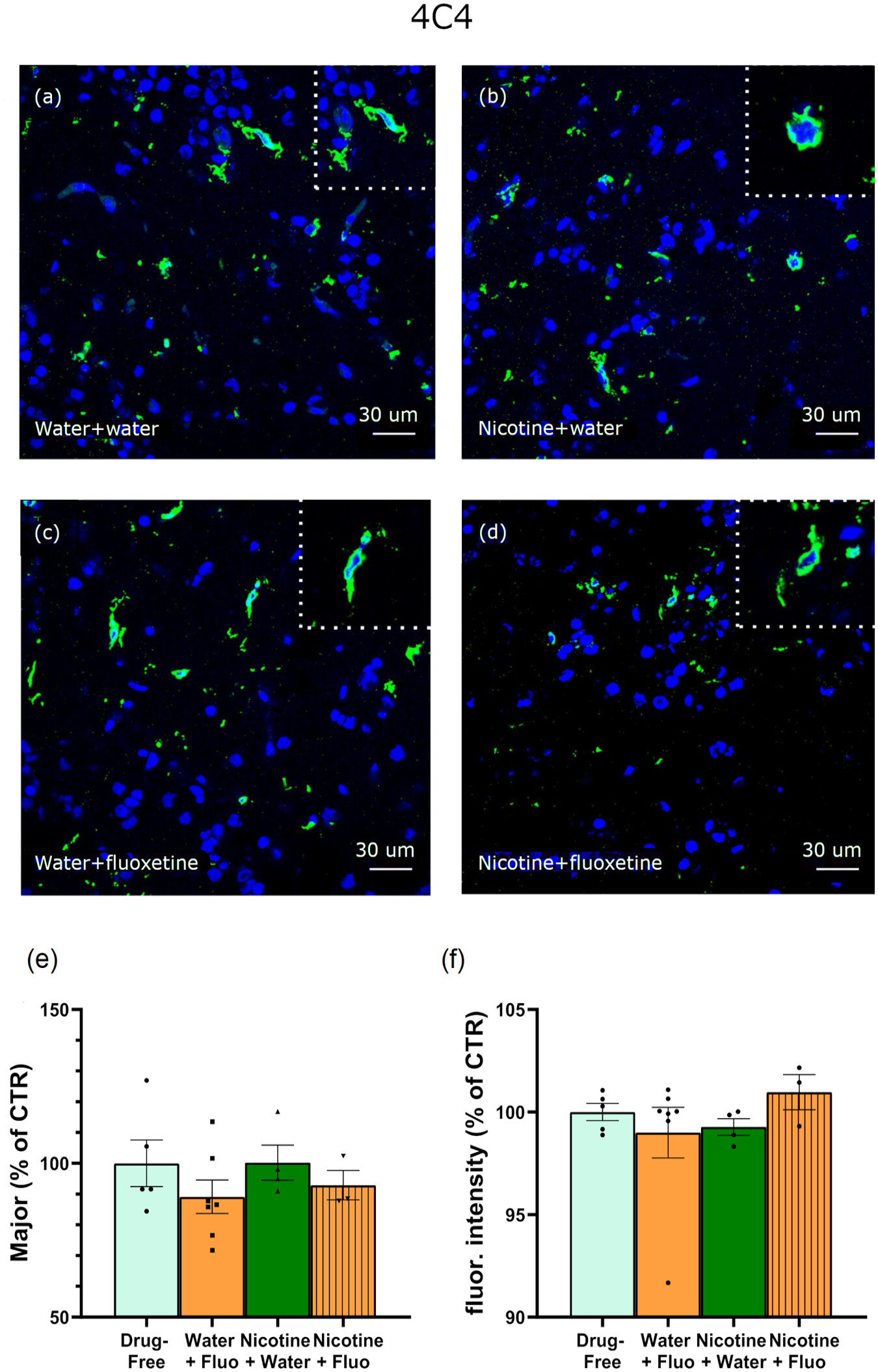
Fig. 8 4C4 expression was evaluated in the PPN 30 days after spontaneous nicotine withdrawal. Representative images obtained from coronal sections show 4C4 immunofluorescence in (a) control (drug-free water + water), (b) nicotine + water, (c) water + fluoxetine and (d) nicotine + fluoxetine. Insets show representative examples of microglial cells. (e, f) Quantitative analysis. No change in 4C4 immunofluorescence area (e) nor in morphological parameters (f) are shown (here major is shown: the major axis of the best fitting ellipse of the analysed cell) (e) nor in 4C4 fluorescence intensity (f). Data are mean ± SEM of the mean of 3/7 zebrafish/group (25?88 cell/group) (one-way analysis of variance (ANOVA)). Each dot represents one animal. (one-way ANOVA).
Image
Figure Caption
Acknowledgments
This image is the copyrighted work of the attributed author or publisher, and
ZFIN has permission only to display this image to its users.
Additional permissions should be obtained from the applicable author or publisher of the image.
Full text @ J. Psychopharmacol. (Oxford)

