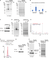
Fig. 4 Znf598 promotes ribosome ubiquitination during development. (A) Comparison of ribosome ubiquitination levels of rpl36-FLAG and MZznf598;rpl36-FLAG embryos. FLAG-IPs from 0 and 24 hpf embryos were subjected to immunoblotting analysis with an anti-Ubiquitin antibody (left) and protein staining (right). The developmental time points are indicated above as hpf. (B) A bar graph shows ubiquitination levels relative to that of rpl36-FLAG embryos at 0 hpf. Ubiquitination signals between 25 and 50 kDa in (A, left) were normalized by corresponding protein amounts in (A, right). The average of three independent experiments is indicated. The error bars indicate the standard deviation. (C,D) Comparison of ribosome ubiquitination levels with or without Znf598 overexpression. Total lysates were subjected to immunoblotting analysis of Znf598-Myc and Tubulin (C). FLAG-IPs were subjected to immunoblotting analysis with an anti-Ubiquitin antibody (D, left) and protein staining (D, right). (E) A representative MS/MS spectrum of Rps10/eS10 di-glycyl K139. (F) MS-based quantification of a ubiquitinated peptide of Rps10/eS10 containing di-glycyl K139 under Znf598 OE (red) and control (blue) conditions. (G,H) Detection of Rps10/eS10 ubiquitination. Total lysates were subjected to immunoblotting analysis of Znf598-Myc and Tubulin (G). FLAG-IPs were subjected to immunoblotting analysis with an anti-Rps10 antibody (H, left) and protein staining (H, right). White and black arrowheads indicate nonubiquitinated or ubiquitinated Rps10/eS10 signals, respectively. The asterisk indicates a nonspecific signal.
Image
Figure Caption
Acknowledgments
This image is the copyrighted work of the attributed author or publisher, and
ZFIN has permission only to display this image to its users.
Additional permissions should be obtained from the applicable author or publisher of the image.
Full text @ RNA

