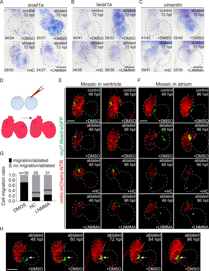
NO signaling regulates CM migration during ventricle regeneration. A–C Whole-mount in situ hybridizations showed reduced expression of snail1a, twist1a and vimentin at 72 hpt in control and ablated hearts treated with DMSO, HC-067047 or L-NMMA for 48–72 hpt. Numbers indicate the ratio of representative staining observed. D–F Injection of myl7:lifeact-eGFP plasmid into Tg(vmhc:mCherry-NTR) embryos at the one-cell stage to monitor the migratory behavior of single eGFP+ CMs in the ventricles or atriums of ablated hearts treated with DMSO, HC-067047 or L-NMMA for 48–72 hpt. G Quantification of the cell migration rate at 96 hpt in ablated hearts treated with DMSO, HC-067047 or L-NMMA for 48–72 hpt. The numbers of larvae analyzed for each condition are indicated. Binomial test, ****P < 0.0001. H Time series images showed the migration process of an eGFP+ CM in the ablated heart at 48–96 hpt. Scale bars, 50 μm. Dashed lines outline the hearts. hpt hours post treatment, CM cardiomyocyte, V ventricle, A atrium, HC HC-067047
Acknowledgments
This image is the copyrighted work of the attributed author or publisher, and
ZFIN has permission only to display this image to its users.
Additional permissions should be obtained from the applicable author or publisher of the image.
Full text @ Cell. Mol. Life Sci.
Your Input Welcome
Thank you for submitting comments. Your input has been emailed to ZFIN curators who may contact you if
additional information is required.
Oops. Something went wrong. Please try again later.