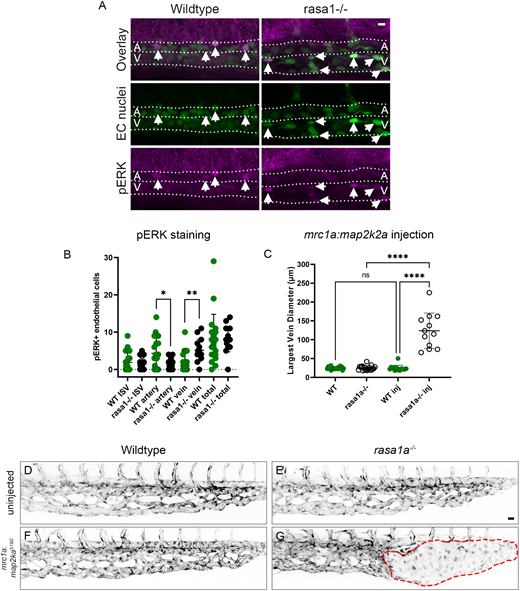
Fig. 7 Ectopic venous activation of pERK in rasa1 mutants drives AVM development. (A) pERK antibody staining in wild type and mutants at 30 hpf. (B) rasa1 mutants show a significant increase in pERK in the vein (wild type, 2.0 cells, rasa1?/?, 5.1 cells; P=0.0020) and a decrease in the DA (wild type, 4.3 cells; rasa1?/?, 1.6 cells; P=0.03) with no change in ISVs at 30 hpf [wild type (n=20), 1.9 cells; rasa1?/? (n=15), 1.4 cells; P=0.5; N=3 experiments, unpaired t-tests]. (C-G) Overexpression of constitutively active map2k2a under a venous promoter (mrc1a) in sensitized rasa1a?/? mutants drives AVM formation. (C) Quantification of largest vein diameter with the ectopic expression of activated mrc1a:map2k2aS219D at 48 hpf. (D,E) Confocal images of uninjected wild type and rasa1a?/? [WTuninj (n=20) versus rasa1a?/?uninj (n=19); P>0.99]. (F,G) Confocal images of wild-type embryos and rasa1a?/? injected with mrc1a:map2k2aS219D [WTinj (n=15) versus rasa1a?/?inj (n=12); P<0.0001; N=2 experiments]. Statistical analysis was carried out using one-way ANOVA with Sidak's correction. Data are meanħs.d. Scale bars: 10 µm in A; 20 µm in D-G.
Image
Figure Caption
Acknowledgments
This image is the copyrighted work of the attributed author or publisher, and
ZFIN has permission only to display this image to its users.
Additional permissions should be obtained from the applicable author or publisher of the image.
Full text @ Development

