
|
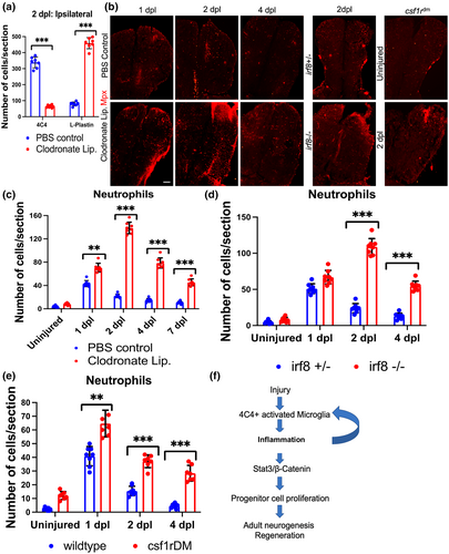
Fig. 8 Persistence of neutrophils when microglia are ablated in the setting of brain injury. (a) Quantification of microglia (4C4) and leukocytes (L-Plastin) in the ipsilesional hemi-telencephala of control or clodronate liposome injected fish at 2 dpl. (b) Confocal images of fish telencephala from PBS control and clodronate treated groups at 1?4 dpl, and irf8+/? (control), irf8?/? and csf1rDM groups at 2 dpl, all immunolabeled for an Mpx-mcherry reporter (neutrophils). (c?e) Quantification of Mpx reporter-positive neutrophils from control, clodronate treated, irf8 and csf1rDM mutant brains. (f) Proposed model of the microglia-induced signaling pathway during injury-induced telencephalic regeneration in zebrafish. ***p < .0001; **p < .001; *p < .01 by t-test for panels (a, c, d, and e). Scale bars = 100 ?m, and the error bars indicate SD.