IMAGE

Fig. 4.

ID
ZDB-IMAGE-231220-64
Source
Figures for Nagorska et al., 2023
Image
Figure Caption

Fig. 4.

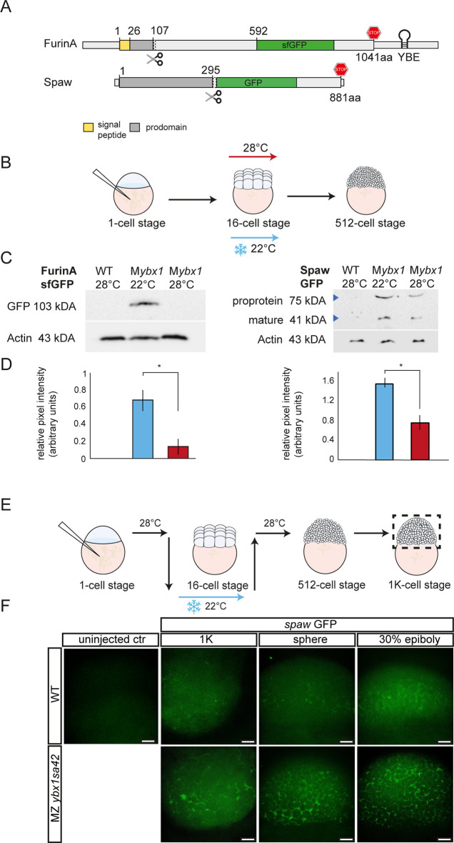
FurinA translation and Spaw maturation is increased in ybx1 mutant embryos. (A) Schematic of GFP fusion reporters for furina X1 and spaw. Hairpin indicates the YBE motif in furina RNA. The FurinA pro-domain is shown in dark grey and the signal peptide in yellow; for Spaw, the pro-domain is indicated in dark grey; stop codon indicated in red in FurinA and Spaw. (B) Schematic of temperature shift of ybx1sa42 mutant embryos. Embryos were injected with GFP reporter-fusion mRNA, grown at 28°C until the 16-cell stage, shifted to 22°C until the 512-cell stage, when protein was extracted. (C) Western blots of embryonic lysates showing FurinA-sfGFP (left) and Spaw-GFP fusion protein (right), following injection of embryos with mRNA reporter. Lanes were loaded with lysates from un-injected wild-type control, Mybx1 at 22°C and Mybx1 control embryos at 28°C. Actin loading control is aligned to the corresponding samples. (D) Bar graphs show quantification of corresponding band intensities, with FurinA-sfGFP (left) and mature Spaw (right) normalised to the Actin loading control, *P<0.05 (P=0.021 for FurinA-GFP; P=0.026 for Spaw-GFP). (E) Schematic showing temperature shift of ybx1 mutant embryos following injection with spaw GFP reporter. (F) Confocal images of 1 K, sphere and 30% epiboly wild-type (top) and ybx1 mutant (bottom) embryos. WT, wild type. Scale bars: 50 µm.

Acknowledgments
This image is the copyrighted work of the attributed author or publisher, and ZFIN has permission only to display this image to its users. Additional permissions should be obtained from the applicable author or publisher of the image. Full text @ Development