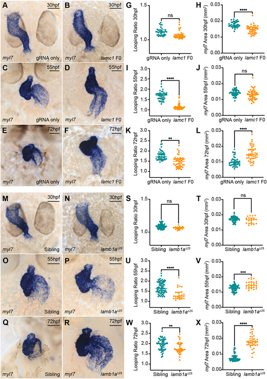
Fig. 2 View largeDownload slide Laminins perform multiple roles during zebrafish heart morphogenesis. (A-F) mRNA in situ hybridisation analysis of myl7 expression in control embryos injected with lamc1-targeting gRNAs only (A,C,E) or with lamc1-targeting gRNAs together with Cas9 protein (lamc1 F0; B,D,F) at 30 hpf (A,B), 55 hpf (C,D) and 72 hpf (E,F). (G-L) Quantitative analysis of looping ratio (G,I,K) and myl7 area (H,J,L) in gRNA-injected controls (30 hpf: n=34; 55 hpf: n=44; 72 hpf: n=44) and lamc1 F0 crispants (30 hpf: n=38; 55 hpf: n=47; 72 hpf: n=44). lamc1 crispants exhibit reduced heart looping at 55 hpf and 72 hpf, a reduced area of myl7 expression at 30 hpf and an increased area of myl7 expression at 72 hpf. Data are median±interquartile range, analysed with the Kruskal?Wallis test. (M-R) mRNA in situ hybridisation analysis of myl7 expression in siblings (M,O,Q) and lamb1a?25 mutants (N,P,R) at 30 hpf, 55 hpf and 72 hpf. (S-X) Quantitative analysis of looping ratio (S,U,W) and myl7 area (T,V,X) in siblings (30 hpf: n=65; 55 hpf: n=70; 72 hpf: n=56) and lamb1a?25 mutants (30 hpf: n=20; 55 hpf: n=25; 72 hpf: n=34). lamb1a?25 mutants exhibit a mild reduction in heart looping from 55 hpf, and an increased area of myl7 expression at 55 hpf and 72 hpf. Data are median±interquartile range, S-W were analysed with the Mann?Whitney U test, X was analysed with the Kruskal-Wallis test. ****P<0.0001, ***P<0.001, **P<0.01, ns=not significant in all graphs. Scale bars: 50 ?m.
Image
Figure Caption
Figure Data
Acknowledgments
This image is the copyrighted work of the attributed author or publisher, and
ZFIN has permission only to display this image to its users.
Additional permissions should be obtained from the applicable author or publisher of the image.
Full text @ Development

