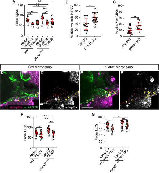
Fig. 5 plxnd1 mutants have elevated Vegfr/Erk signalling in the facial lymphatics. (A) Quantitation of facial LEC number in 6 dpf larvae treated from 3 to 6 dpf with either DMSO, 1 µM sunitinib or 2 nM tivozanib. (B,C) Quantitation of the proportion of pErk-positive cells within the PCV at 36 hpf (B) or the facial LECs at 3 dpf (C). (D-E′) Confocal images of anti-pErk (magenta) and anti-EGFP (green) staining in the head at 3 dpf of either control (D,D′) or plxnd1 (E,E′) lyve1b:EGFP morphant larvae. (D′,E′) Anti-pErk staining only. Yellow arrowheads indicate the pErk cells within the facial lymphatics. (F) Quantitation of facial LEC number in 6 dpf larvae treated from 3 to 6 dpf with either DMSO or 5 µM SL327. (G) Quantitation of facial LEC number in lyve1b:DsRed; fli1a:nlsEGFP larvae injected with either control or vegfd morpholinos. P>0.05 (not significant), *P<0.05, **P<0.01 (unpaired t-tests, Kruskal–Wallis and ANOVA); data are mean±s.d. Scale bar: 50 µm. LEC, lymphatic endothelial cell. Numbers in graphs represent numbers of larvae.
Image
Figure Caption
Figure Data
Acknowledgments
This image is the copyrighted work of the attributed author or publisher, and
ZFIN has permission only to display this image to its users.
Additional permissions should be obtained from the applicable author or publisher of the image.
Full text @ Development

