IMAGE

Figure 4

ID
ZDB-IMAGE-231204-38
Antibodies
Source
Figures for Serrano et al., 2023
Image
Figure Caption

Figure 4

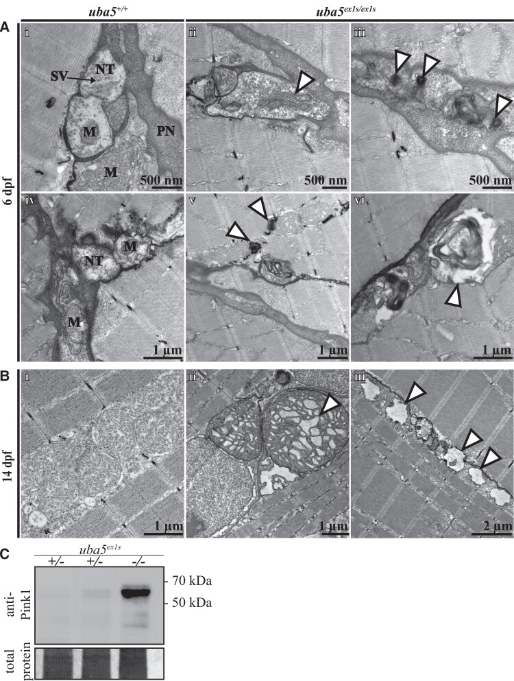
Ultrastructural analysis of the peripheral nerves and skeletal muscle in uba5ex1s/ex1s. (A) Patterning of the peripheral nerves and skeletal muscle at 6 dpf (i–vi). Electron microscopy images show mitochondrial abnormalities, including elongated mitochondria (ii) and degenerating mitochondria (iii), in the nerve terminals of uba5ex1s/ex1s fish (arrowhead). Degradation vacuoles enclosing debris (vi), and autophagic structures associated with mitochondria in the skeletal muscle (v), are also observed. (B) Patterning of the skeletal muscle at 14 dpf shows abnormal mitochondria with swollen cristae (ii) and vacuolated mitochondria (iii) in uba5 mutants (arrowhead). (C) Western blot for Pink1 and Direct Blue staining (loading control) on protein lysates obtained from pooled samples of 25 embryos at 6 dpf. M, mitochondria; NT, nerve terminal; PN, peripheral nerve; SV, synaptic vesicles.

Figure Data
Acknowledgments
This image is the copyrighted work of the attributed author or publisher, and ZFIN has permission only to display this image to its users. Additional permissions should be obtained from the applicable author or publisher of the image. Full text @ Brain Commun