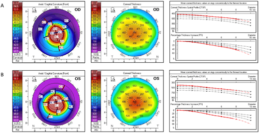
Fig. 1 Clinical ocular features of the ZNF469 mutant patient. (A) Pentacam refractive maps and corneal thickness map of the right eye (OD). Thinnest corneal thickness = 435 Ám, Kmax = 64.9 D. (B) Pentacam refractive maps and corneal thickness map of the left eye (OS). Thinnest corneal thickness = 406 Ám, Kmax = 70.4 D. Pupil center is indicated by a plus sign (+), the pachy apex is marked with a filled circle (?), and the thinnest location is indicated by an open circle (?). Three black dotted lines in the corneal thickness map indicate the distribution of healthy people, and the red line indicates the fitting curve of the patient.
Image
Figure Caption
Acknowledgments
This image is the copyrighted work of the attributed author or publisher, and
ZFIN has permission only to display this image to its users.
Additional permissions should be obtained from the applicable author or publisher of the image.
Full text @ Invest. Ophthalmol. Vis. Sci.

