IMAGE

Figure 2

ID
ZDB-IMAGE-231030-109
Genes
Antibodies
Source
Figures for Huttner et al., 2023
Image
Figure Caption

Figure 2

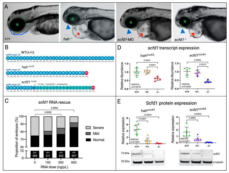
scfd1 deficiency is responsible for the hahvcc43 mutant phenotype. (A) Brightfield photographs show similar phenotypes in hahvcc43−/− mutants (middle left), scfd1 morphants (middle right), and scfd1vcc44−/− mutants (right) in comparison to wildtype (WT; (+/+; left) at 3 days post fertilization (dpf). These include cardiac effusion and poorly ballooned chambers (red arrow), as well as craniofacial defects such as an absence of lower jaw (blue arrowheads) in comparison to a clearly defined jawline in +/+ (blue outline) and small eyes (green asterisk in dotted circle). (B) Both hahvcc43 and scfd1vcc44 mutants harbor a PTC in exon 10 of scfd1. (C) Bar graph showing the proportion of hah embryos exhibiting normal (black bars), mild (dark gray bars), or severe (light gray bars) mutant phenotypes at 4 dpf after injection with WT scfd1 mRNA. Note reduction in proportion of affected embryos with increasing concentrations of scfd1 mRNA. See Methods for definition of phenotypes. Numbers of embryos in each group indicated at base of bar. Chi-square p-values stated. (D,E) Scatter plots showing reduced scfd1 transcript expression as determined by qRT-PCR (D; 30 embryos/sample, n = 5 samples) and protein expression as determined using Western blot (E; 30 embryos/sample, n = 6 samples) in both heterozygous (+/−) and homozygous (−/−) hahvcc43 and scfd1vcc44 mutants at 3 dpf. Ordinary one-way ANOVA p-values stated. Significance if p < 0.05. α-tubulin loading control used for normalization for Western blot quantification. Full blots are shown in Supplementary Figure S2. Created with BioRender.com.

Figure Data
Acknowledgments
This image is the copyrighted work of the attributed author or publisher, and ZFIN has permission only to display this image to its users. Additional permissions should be obtained from the applicable author or publisher of the image. Full text @ J Cardiovasc Dev Dis