IMAGE

Fig. 2.

ID
ZDB-IMAGE-231018-32
Genes
Antibodies
Source
Figures for Ali et al., 2023
Image
Figure Caption

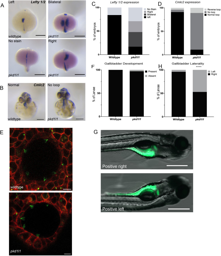
Fig. 2.

pkd1l1 mutant embryos demonstrate laterality defects. (A,B) Whole-mount RNA in-situ hybridization demonstrating lefty 1 and lefty 2 (lefty 1/2) expression in wild-type and pkd1l1 mutant embryos (dorsal view at 19 hpf) (A) and cmlc2 expression in wild-type and pkd1l1 mutant embryos (ventral view at 48 hpf) (B). Scale bars: 0.25 mm. (C) lefty 1/2 expression was only demonstrated on the left side in wild-type embryos (n=45), with expression being dispersed among the left, right and bilateral sides in pkd1l1 mutant embryos (n=48). (D) cmlc2 expression demonstrated normal looping in the majority of wild-type embryos (n=74), but in pkd1l1 mutant embryos (n=36), looping was primarily absent, and a few pkd1l1 mutant embryos showed either normal or reverse looping. (E) Confocal images of the KV of 10-16 somite wild-type (n=5) and pkd1l1 mutant (n=7) embryos demonstrate the presence of motile cilia. Arl-GFP is shown in green and membrane-localized RFP (memRFP) in red. Scale bars: 10 μm. (F) Quantification of the presence of the gallbladder showed no significant differences in gallbladder development in wild-type (n=33) and pkd1l1 mutant (n=39) larvae. (G) Position of the gallbladder as seen from right and left lateral views. Scale bars: 500 µm. (H) pkd1l1 mutations cause laterality defects in gallbladder positioning as demonstrated by an increased frequency of left-sided gallbladders in pkd1l1 mutant (n=156) compared to wild-type (n=166) zebrafish. ****P<0.0001 (Fisher's exact test).

Figure Data
Acknowledgments
This image is the copyrighted work of the attributed author or publisher, and ZFIN has permission only to display this image to its users. Additional permissions should be obtained from the applicable author or publisher of the image. Full text @ Dis. Model. Mech.