IMAGE

Figure 3.

ID
ZDB-IMAGE-231012-9
Source
Figures for Oyarbide et al., 2023
Image
Figure Caption

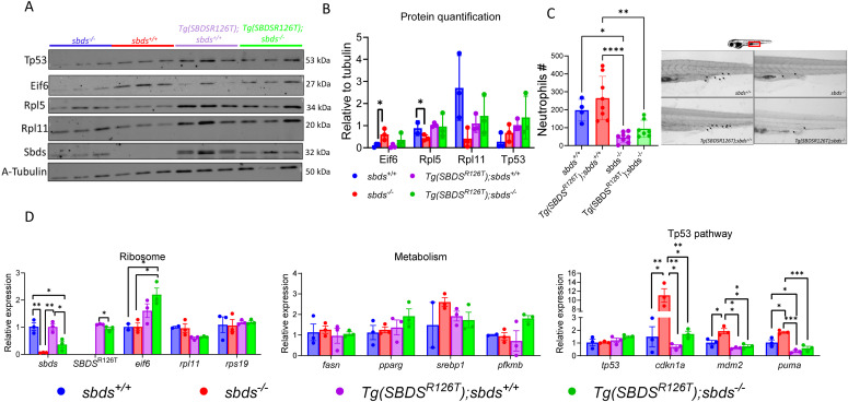
Figure 3. Phenotype of 10 dpf larvae expressing the human SBDSR126T.

(A) Western blot showed Eif6 without RPL5, and Rpl11 accumulation in the sbds KO fish, but there was no Eif6 accumulation in the SBDSR126T strain. (B) Protein quantification of Western blots as performed with NIH ImageJ. (C) After Sudan black staining, neutrophils were counted and found to be significant lower in the sbds KO background with or without the transgene. (D) qRT-PCR analysis of critical genes involved in ribosomal, metabolic, and tp53-associated pathways.

Acknowledgments
This image is the copyrighted work of the attributed author or publisher, and ZFIN has permission only to display this image to its users. Additional permissions should be obtained from the applicable author or publisher of the image. Full text @ Life Sci Alliance