IMAGE

Fig. 3.

ID
ZDB-IMAGE-231002-102
Source
Figures for Leid et al., 2023
Image
Figure Caption

Fig. 3.

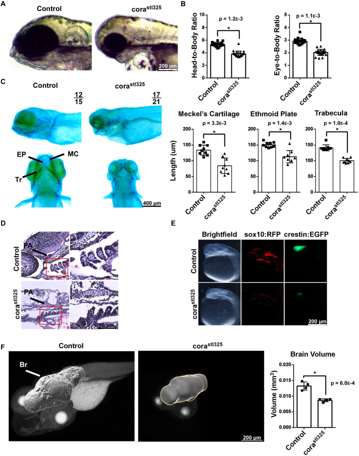
corastl325 embryos have craniofacial deformities and neuroanatomical defects. (A) Head morphology of control and corastl325 embryos at 96 hpf. (B) Head-to-body and head-to-eye ratios. Control (n=13), corastl325 (n=9). (C) Alcian Blue staining of control and corastl325 embryos at 96 hpf (left), with quantification of major jaw elements (right). EP, Ethmoid plate; MC, Meckel's Cartilage; T, Trabeculae. n=9 per experimental group. (D) H&E staining of the jaw of control and corastl325 embryos at 96 hpf. (E) Fluorescence microscopy images of Tg(crestin::GFP) and Tg(sox10::RFP) control and corastl325 embryos. (F) X-ray microscopy-generated images of 96 hpf embryos (left) and quantification of tissue volume (right). Br, brain Control (n=4), corastl325 (n=4).

Figure Data
Acknowledgments
This image is the copyrighted work of the attributed author or publisher, and ZFIN has permission only to display this image to its users. Additional permissions should be obtained from the applicable author or publisher of the image. Full text @ Biol. Open