Fig. 4
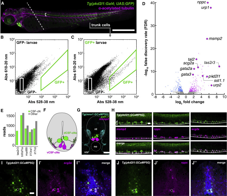
Figure 4. The CSF-cN transcriptome reveals the expression of taste receptors and immune-related secreted factors (A) 3 dpf zebrafish Tg(pkd2l1:GAL4; UAS:GFP) larva immunostained for GFP (green) and acetylated tubulin (magenta). Dashed line indicates plane of decapitation prior to dissociation (anterior tissues were discarded to exclude labeled cells in brain and heart); bracket indicates CSF-cN domain. Scale bars, 500 ?m. (B) Calibration FACS plot from non-transgenic siblings. (C) FACS plot from a typical dissociation of cells from transgenic larvae. A small fraction (?0.2% of input) of cells is green-shifted. Figure S3 quantifies the upper bound of cells/larvae we could reasonably expect to obtain. In Figure S4A, we use qPCR to show that this fraction appeared to be CSF-cNs. (D) Volcano plot of all RNA-seq replicates: 202 transcripts were enriched in CSF-cNs. Figure S4B provides more categorical information on these hits. Figure S6 shows that knocking out individual CSF-cN-enriched taste receptors identified by this approach did not impair CSF-cNs? ability to detect bitter compounds. (E) Quantification of reads for selected immune-related receptors in both GFP+ CSF-cNs and GFP? (i.e., all other) cells. (F) Schematic representation of a transverse section of the spinal cord and central canal showing the disposition of dorsolateral CSF-cNs (dCSF-cNs, green) and ventromedial CSF-cNs (vCSF-cNs, magenta). (G) A mixed fluorescence in situ hybridization/immunofluorescent stain showing the disposition of motor neurons (cyan, Tg(mnx1:GCaMP5)), dCSF-cNs (green, sst1.1), and vCSF-cNs (magenta, msmp2). The section here is a frontal section, e.g., a cross-section of the spinal cord. Scale bars, 50 ?m. (H) Fluorescence in situ hybridization reveals that the putative immune-related secreted factors msmp2, nppc, and scg2a are expressed in CSF-cNs. These confocal stacks are lateral stacks, so at 90° orientation to the image in (G). Scale bars, 50 ?m. Figure S5 shows additional in situ hybridization results from some of these RNA-seq hits; most hits we assessed were verified histologically. (I) Transverse section of adult Tg(pkd2l1:GCaMP5G) spinal cord; expression of scg2a is specific to CSF-cNs. Scale bars, 50 ?m. (J) Adults continue to express nppc and scg2a in CSF-cNs. See also Figures S3?S6, Table S1, and Data S1.
Image
Figure Caption
Acknowledgments
This image is the copyrighted work of the attributed author or publisher, and
ZFIN has permission only to display this image to its users.
Additional permissions should be obtained from the applicable author or publisher of the image.
Full text @ Curr. Biol.

