Fig. 2
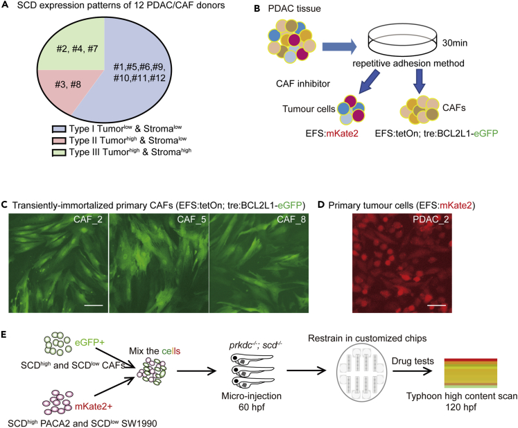
Harvest of primary PDAC tumor cells and CAFs for zPDX models
(A) Type proportions of the twelve stroma-enriched surgical-resected PDAC tissues from local hospital.
(B) Schematic procedure to isolate CAFs and tumor cells from primary PDAC tissues.
(C) Representative fluorescent images of the primary CAFs transfected by lentivirus (EFS: BCL2L1-P2A-eGFP).
(D) Representative fluorescent images of the primary tumor cells transfected by lentivirus (EFS: mKate2).
(E) Schematic procedure to generate the stromal-tumor-mixed zPDX models in scd−/−/prkdc−/− zebrafish larvae and alignment of the zPDX models in a microarray chip. Scale bar: 20 μm.

