
|
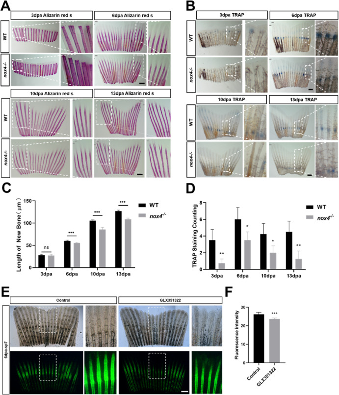
Fig. 5 Fig. 5. Nox4 loss reduces tail regeneration. (A) Alizarin red and (B) TRAP staining were performed 3, 6, 10, and 13 days postamputation (dpa). The figures show the front view and an enlarged view of the tail. (C) Regeneration length in WT and nox4 −/− tails at different growth stages. (D) Quantification of TRAP staining in WT and nox4 −/− zebrafish tail at different growth stages. (E) Tg(sp7-EGFP) zebrafish were treated with GLX351322 and fluorescence was observed at 6 dpa. The panels show the front view and an enlarged view of the tail. (F) Quantification of green fluorescence in osteoblasts. n = 6. The scale bars in panels A, B, E are 200 μm.The results were analyzed using one-way ANOVA. *P < 0.05, **P < 0.01, and ***P < 0.001. (For interpretation of the references to colour in this figure legend, the reader is referred to the Web version of this article.)
Reprinted from Free radical biology & medicine, 193(Pt 2), Cao, Z., Liu, G., Zhang, H., Wang, M., Xu, Y., Nox4 promotes osteoblast differentiation through TGF-beta signal pathway, 595-609, Copyright (2022) with permission from Elsevier. Full text @ Free Radic. Biol. Med.