Figure 4
Figure 4
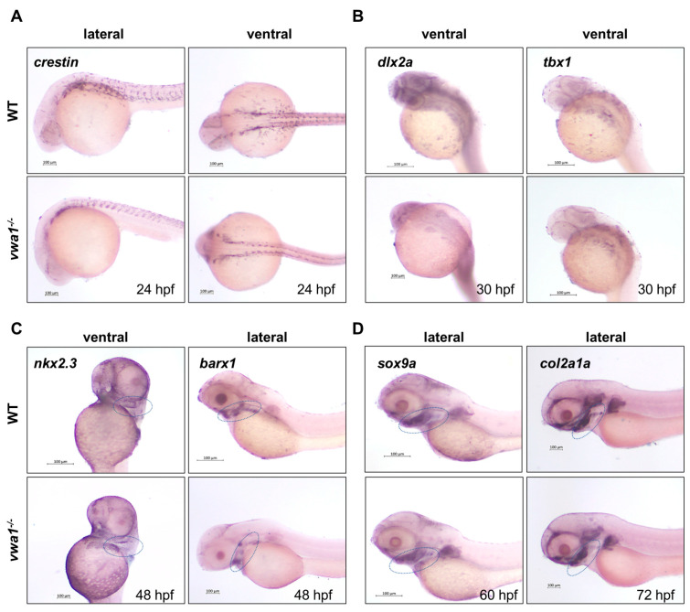
vwa1 functions in cranial neural crest cell condensation. (A) WISH analysis of crestin at 24 hpf showed no significant difference between vwa1 mutants and WT controls. (B) The expression pattern of dlx2a and tbx1 were essentially normal in the pharyngeal arch at 30 hpf, although the absolute area was smaller owing to the reduced body size of mutants. (C) At 48 hpf, expression of the endodermal pouch marker, nkx2.3, was not significantly different between mutant embryos and WT controls. However, the expression of barx1, indicative of the condensation of prechondral mesenchymal cells, was significantly reduced in the pharyngeal region of vwa1 mutants compared with that in WT controls. (D) Expression level of sox9a did not change significantly at 60 hpf, whereas the expression of col2a1a decreased at 72 hpf. The mandibular region is marked with dotted blue circles.

