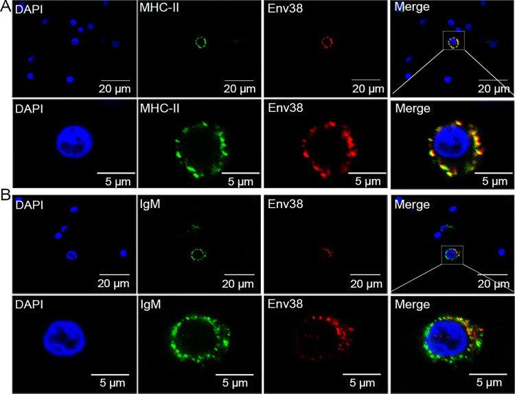
Fig 4
The leukocytes were sorted from spleen, head kidney and peripheral blood of zebrafish stimulated with SVCV (105 TCID50). (A) Cells were labled with mouse anti-Env38 and rabbit anti-MHC-II? Abs, followed by Alexa Fluor 594-conjugated goat anti-mouse IgG and FITC-conjugated goat anti-rabbit IgG. (B) Cells were labled with mouse anti-Env38 and rabbit anti-IgM Abs, followed by Alexa Fluor 594-conjugated goat anti-mouse IgG and FITC-conjugated goat anti-rabbit IgG. DAPI stain showed the location of the nuclei. Fluorescence images were captured using a Laser scanning confocal microscope (FV3000) with 60 × oil glass.

