
|
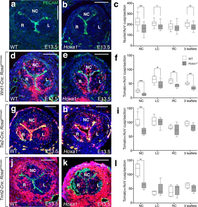
Fig. 5
a, b Transversal sections of E13.5 hearts immunostained with an endothelial marker (anti-Pecam antibody, green) and DAPI staining for nuclei (blue). Note the smaller non-coronary (NC) leaflet in Hoxa1−/− (b) compared with WT (a) embryos. c Total nuclei were counted in the three forming aortic valve leaflets from Hoxa1−/− (n = 20) and WT (n = 18) embryos at E13.5 spanning a 180 μm depth (***p = 0.0006; **p = 0.0009; *p = 0.01). d–l Fate-mapping of the neural crest-, endothelial- and second heart field-lineages in the aortic valves of WT (d, g, j), and Hoxa1-null (e, h, k) embryos at E13.5. Tomato-reporter is visualized in red and nuclei (DAPI) appear in blue.