Image
Figure Caption
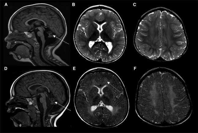
Fig. 1 MRI characteristics of individual 1
Brain MRI of individual 1 at 17 months (A?C) and 35 months of age (D?F).
(A) and (D) Sagittal T1-weighted images at the midline showing cerebellar hypoplasia (thin arrows).
(B, C, E, and F) Axial T2-weighted images at the level of the basal ganglia (B and E) and centrum semiovale (C and F) showing progressive diffuse demyelination (dotted arrow).
Acknowledgments
This image is the copyrighted work of the attributed author or publisher, and
ZFIN has permission only to display this image to its users.
Additional permissions should be obtained from the applicable author or publisher of the image.
Full text @ HGG Adv

