Image
Figure Caption
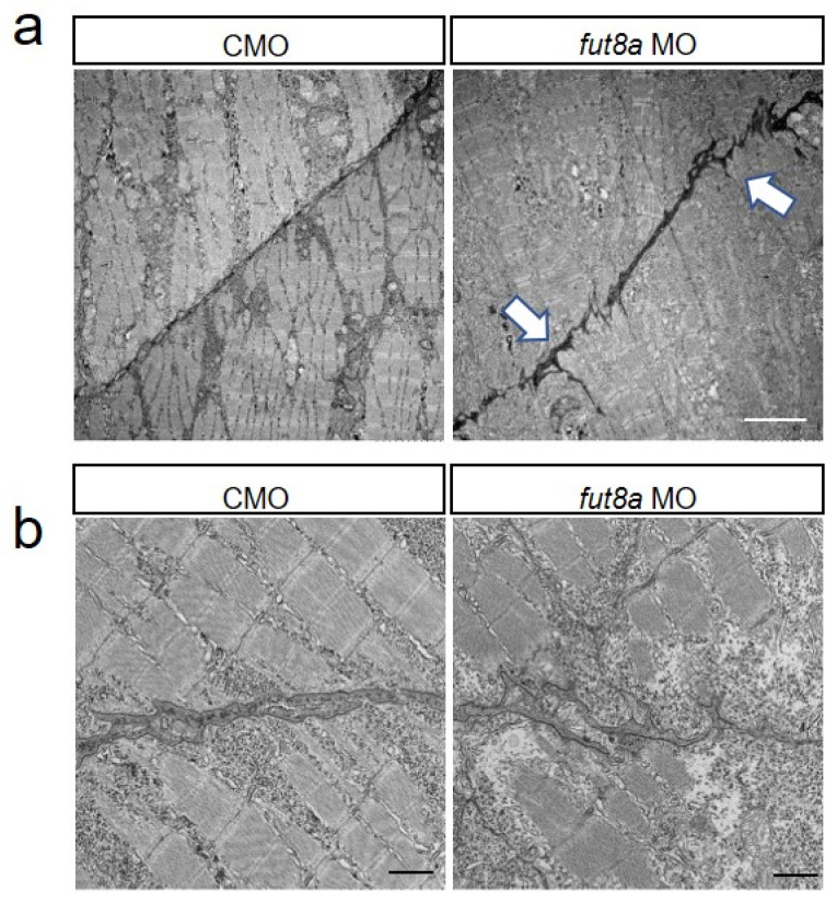
Figure 4 fut8a morphants have abnormal myosepta Representative electron micrographs of longitudinal section of morphants (2 dpf). (a) Electron micrograph of dorsal myosepta. White arrows indicate the tear sites in the myosepta. Scale bar: 5 μm. (b) Enlarged views of electron micrograph of dorsal myosepta. Scale bar: 1 μm. Maximum width, total length, and straightness factor of myosepta were quantified and indicated in Supplemental Figure S2.
Figure Data
Acknowledgments
This image is the copyrighted work of the attributed author or publisher, and
ZFIN has permission only to display this image to its users.
Additional permissions should be obtained from the applicable author or publisher of the image.
Full text @ Cells

