
|
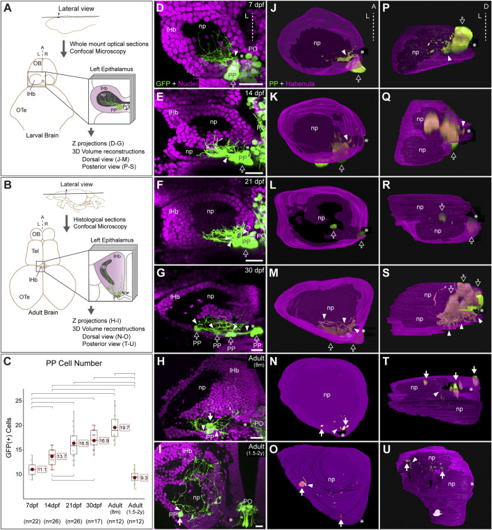
FIGURE 1 Post-hatching ontogeny of parapineal morphology and connectivity in zebrafish. (A,B) Schemes of larval (A) and adult (B) zebrafish brains showing the overall anatomy of the epithalamus in dorsal views and the position of optical and histological sections analysed in the rest of the panels. The left side of the epithalamus is highlighted and enlarged in a square containing the left habenula (magenta) and the parapineal (green). The position of the habenular commissure is indicated with an asterisk. (C) Box plot showing the quantification of GFP(+) parapineal cells in larvae (7, 14 and 21 dpf), juveniles (30 dpf) and adults (8 mpf and 1.5?2 years old). N for each group are shown below the x-axis. Significance was set to p < 0.001 (upper square brackets) or p < 0.01 (lower square brackets). The red circle on each bar is the mean for the group and its value is indicated with red square labels. The horizontal line in the box is the median of the group. (D?I) Ontogeny of parapineal morphology and connectivity revealed by immunofluorescence against GFP (green) and DAPI/Hoechst staining (magenta) in Tg(foxd3::GFP) zebrafish. Images correspond to dorsal views of representative confocal z-stacks maximum projections of the left epithalamus at 7 dpf (D), 14 dpf (E), 21 dpf (F), 30 dpf (G), 8 months (H) and 1.5?2 years old (I), with anterior to the top, left to the left and the midline (dotted line) towards the right side of the panel. Arrowheads indicates the site where parapineal projections emerge, often forming a thick bundle at earlier stages. Empty arrows indicate the parapineal cell body as long it is recognizable as a single large cluster. White arrows indicate parapineal cell groups scattered along the posterior border of the left habenula. (J?O) Dorsal views of 3D reconstructions of the left habenula (magenta = cell bodies; black = neuropil) and parapineal (green) at 7 dpf (J), 14 dpf (K), 21 dpf (L), 30 dpf (M), 8 months (N) and 1.5?2 years old (O), with anterior to the top, left to the left and the midline (dotted line) towards the right side of the panel. (P?U) Posterior views of 3D reconstructions of the left habenula (magenta = cell bodies; black = neuropil) and parapineal (green) at 7 dpf (P), 14 dpf (Q), 21 dpf (R), 30 dpf (S), 8 months (T) and 1.5?2 years old (U), with dorsal to the top, left to the left and the midline (dotted line) towards the right side of the panel. Abbreviations: A (anterior), D (Dorsal), L (left), lHb (left habenula), np (habenular neuropil), OB (Olfatory Bulb), OTe (Optic Tectum), PO (pineal organ at the level of the stalk), PP (parapineal), R (right), Tel (Telencephalon). Scale bars, 10 �m.