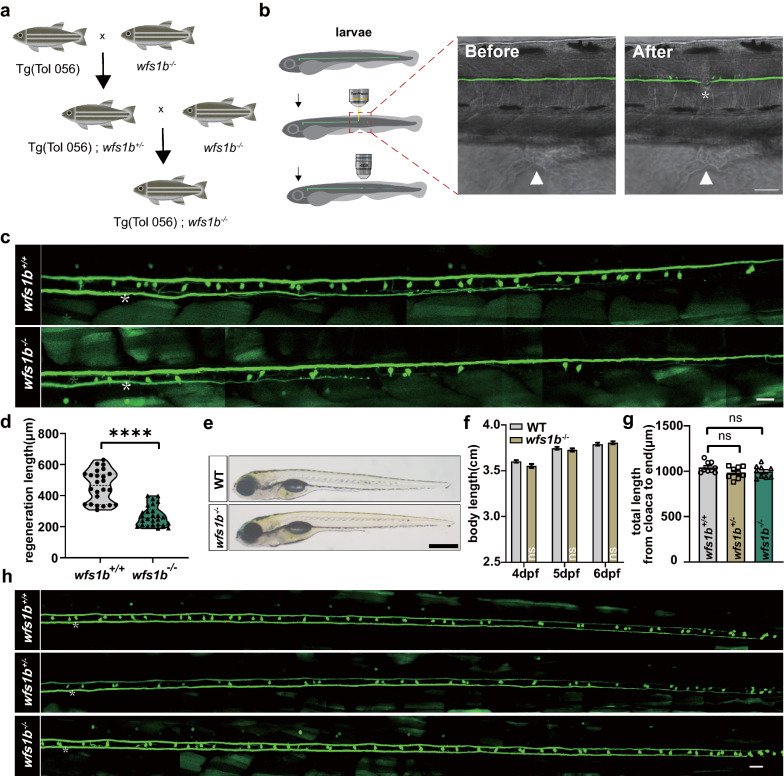
Fig. 3 Wfs1b regulates M-cell axon regeneration in vivo. a Hybridization of the transgenic line: Tg (Tol 056: EGFP) and wfs1b mutants were crossed for two consecutive generations to obtain Tg (Tol 056: EGFP)/ wfs1b +/- and Tg (T056: EGFP); wfs1b ?/? lines. b Representative images of the M-cell axon before and after ablations by a two-photon laser. Asterisk, injury site; arrowhead, cloacal pore; scale bar, 50 ?m. c, d Confocal imaging of M-cell axons between wfs1b+/+ and wfs1b?/? groups at 8 dpf and the regeneration length at 2 dpa. Violin plot shows all data points, including minimum, maximum, median, and quartiles. Scale bar, 20 ?m, P < 0.0001, control, n = 24; wfs1b?/?, n = 26. Assessed by unpaired t test. e, f Representative images of embryos from the wildtype and the mutant at 6 dpf (scale bar, 500 ?m), and measured total body length from 4 to 6 dpf before axotomy (4 dpf, wildtype: 3.603 ± 0.01402 cm, wfs1?/?: 3.549 ± 0.02201 cm, P = 0.1708; 5 dpf, wildtype: 3.745 ± 0.01504 cm, wfs1?/?: 3.727 ± 0.01679 cm, P = 0.8255; 6 dpf, wildtype: 3.790 ± 0.01691 cm, wfs1?/?: 3.806 ± 0.01569 cm, P = 0.8842; n = 30). Assessed by two-way ANOVA. ns, not significant. g, h Defined lengths of M-cell axons from the cloaca to the end were not notably different among WT, homozygous, and heterozygous larvae (wfs1b+/+: 1042 ± 19.51 ?m; wfs1b+/-: 985.2 ± 21.47 ?m, P = 0.0689; wfs1b?/?: 995.6 ± 21.98 ?m, P = 0.1363; n = 9). Assessed by ordinary one-way ANOVA/Tukey?s multiple-comparisons test (wfs1b+/+ versus wfs1b+/-: P = 0.1594; wfs1b+/+ versus wfs1b?/?: P = 0.2856; wfs1b+/- versus wfs1b?/?: P = 0.9342) White asterisk: ablation point. Scale bar, 20 ?m. ns, not significant
Image
Figure Caption
Figure Data
Acknowledgments
This image is the copyrighted work of the attributed author or publisher, and
ZFIN has permission only to display this image to its users.
Additional permissions should be obtained from the applicable author or publisher of the image.
Full text @ Acta Neuropathol Commun

