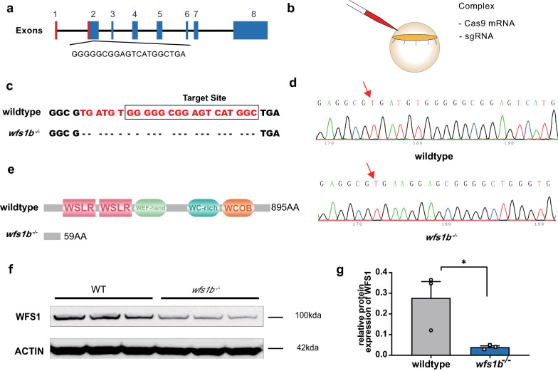
Fig. 2 Generation and identification of wfs1b mutant zebrafish. a Schematic of the Cas9-sgRNA targeted site located at the second exon of wfs1b. b schematic of the complex injected into one-cell embryos. c, d Representative sequencing results of wide-type and mutated zebrafish lines. The mutant sequencing result showed a 23-bp base deletion. e Bioinformatics analysis indicated that the mutated region is located in the front of the functional domain of Wfs1b. The wildtype translated to 895 amino acids, whereas the mutant translated to 59 normal amino acids. f, g Western-blotting analysis showed that WFS1 protein expression is inhibited in the mutant group compared with that of the wildtype. The protein expressions were quantified by Image J software. The experiment was repeated three times with three independent samples. P = 0.0394. Assessed by unpaired t test
Image
Figure Caption
Acknowledgments
This image is the copyrighted work of the attributed author or publisher, and
ZFIN has permission only to display this image to its users.
Additional permissions should be obtained from the applicable author or publisher of the image.
Full text @ Acta Neuropathol Commun

