
|
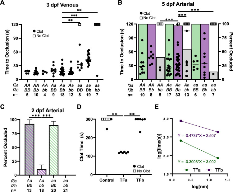
Fig 4
(A) Laser-mediated venous endothelial injury at 3 dpf shows that the time to occlusion (TTO) was not altered by loss of TFb (AA/bb, Aa/bb) but was delayed in the complete absence of TFa (aa/BB, aa/Bb). Thrombus formation was absent after complete TF loss (aa/bb). (B) Laser-mediated arterial endothelial injury at 5 dpf shows that the ability to form occlusive thrombi was significantly reduced in fish lacking TFb (AA/bb, Aa/bb, aa/bb, indicated by grey bars) compared to BB and Bb groups (green and purple bars; respectively). (C) Endothelial injury at 2 dpf confirms the dependence on TFb in the arterial system prior to the presence of thrombocytes. Occlusive thrombi were nearly absent without TFb (Aa/bb, aa/bb). (D) Relipidated recombinant TFa or TFb proteins (173 nM) were mixed with citrated trout plasma, and recalcified. TFa-liposomes induced clot formation during a manual tilt test significantly faster than TFb-liposomes or empty liposome controls. The reaction was observed until 300 seconds, and then again at 15 minutes to assess for delayed clot formation (the latter observation is indicated by an open or closed circle at 300 seconds). (E) The assay was repeated with relipidated TF diluted in 0.5x HBS (20 mM HEPES pH 7.5, 50 mM NaCl) and plotted on log-log axes demonstrating 10 to 100-fold higher procoagulant activity of TFa over TFb. Statistical significance by Mann-Whitney U testing: ** < 0.01, *** < 0.001. All phenotypic data were collected by an observer blinded to genotype. Open squares in A and B indicate individual larvae that did not occlude within 120 seconds.