Image
Figure Caption
Figure 2
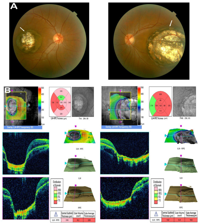
Clinical observations. (A) Fundus photograph of the proband (II:3) exhibiting prominent bilateral atrophy in the macula (white arrows); the atrophy presents well-circumscribed borders, total chorioretinal maldevelopment with a white appearance, and increased backscatter around the lesion due to the bare sclera. (B) OCT scan of the proband (II:3) showing a cup-shaped lesion with the complete absence of retina and choroid. There is a sharp margin, with significant excavation in the foveal region of both eyes.
Acknowledgments
This image is the copyrighted work of the attributed author or publisher, and
ZFIN has permission only to display this image to its users.
Additional permissions should be obtained from the applicable author or publisher of the image.
Full text @ Cells

