Image
Figure Caption
Fig. 6
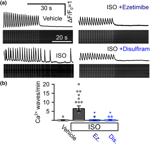
Suppression of arrhythmogenic Ca2+ signals in iPSC-derived cardiomyocytes from a catecholaminergic polymorphic ventricular tachycardia (CPVT) patient. (a) Representative confocal linescan recordings of intracellular Ca2+ in iPSC-derived cardiomyocytes from a CPVT patient. Cells were continuously pulsed at 0.5 Hz and spontaneous Ca2+ waves were analysed after pulsing was stopped. Addition of isoprenaline (ISO) induced spontaneous Ca2+ waves which could be blocked by the addition of ezetimibe or disulfiram. (b) Quantitative analysis of the experiments in (a). Addition of ISO induced the occurrence of waves in CPVT cells (0 waves per minute for vehicle control, n = 11 cells, 5.87 ± 1.80 under ISO, n = 16, Kruskal?Wallis test), which could be suppressed by the addition of ezetimibe (Ez) or disulfiram (Dis) (0.10 ± 0.07 waves per minute for ezetimibe, n = 13 cells, 0.05 ± 0.05 waves per minute for disulfiram, n = 13, Kruskal?Wallis test, data from four independent enzymatic dissociations of 19 individual beating cell clusters)
Acknowledgments
This image is the copyrighted work of the attributed author or publisher, and
ZFIN has permission only to display this image to its users.
Additional permissions should be obtained from the applicable author or publisher of the image.
Full text @ Br. J. Pharmacol.

