
|
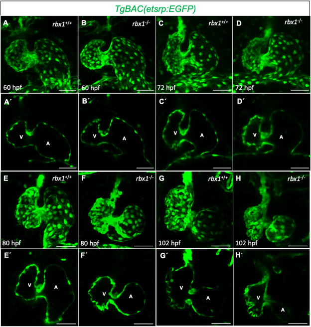
Fig. 3 The endocardium is affected in rbx1 mutants
(A-H?) 3D Confocal images (maximum intensity projections) (A?H), and 2D mid-sagittal sections (A?-H?) of the endocardium from rbx1 animals. (A-F?) Endocardial morphology in rbx1+/+ and rbx1?/? siblings at 60 (A-B?), 72 (C-D?) and 80 (E-F?) hpf. At 72 hpf a minor reduction in ventricular size is visible in rbx1 mutants (C-D?). At 80 hpf, rbx1 mutants exhibit a smaller ventricle with a stretched AV canal and outflow tract (E-F?). (G-H?) Endocardial morphology in rbx1+/+ (G-G?) and rbx1?/? (H?H?) siblings at 102 hpf. rbx1?/? larvae exhibit a smaller ventricle (H?H?) compared with rbx1+/+ (G-G?) siblings.
Reprinted from Developmental Biology, 480, Sarvari, P., Rasouli, S.J., Allanki, S., Stone, O.A., Sokol, A., Graumann, J., Stainier, D.Y.R., The E3 ubiquitin-protein ligase Rbx1 regulates cardiac wall morphogenesis in zebrafish, 1-12, Copyright (2021) with permission from Elsevier. Full text @ Dev. Biol.