Fig 5
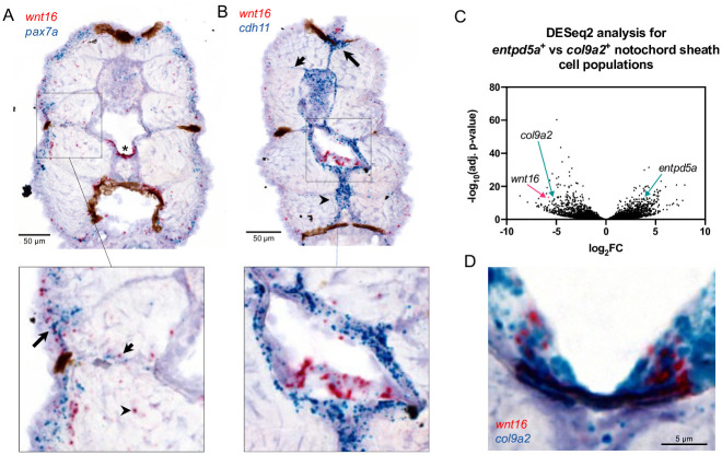
(A, B) Chromogenic in situ hybridization was performed in transverse sections through the zebrafish trunk at 12 dpf. (A) Co-staining for wnt16 and pax7. Staining for wnt16 was detected in notochord (star) and was mostly negative for pax7a. Staining for wnt16 was also detected in cells in the lateral portion of the myotome (inset, long arrow), within or adjacent to presumptive myosepta (inset, short arrow), and sporadically within the myotome (inset, arrowhead). (B) Co-staining for wnt16 and cdh11. Staining for cdh11 was apparent in cells surrounding the notochord, the dorsal myotome (long arrow), presumptive myosepta (short arrow), and the medial portion of the ventral myotome (arrowhead). With regard to notochord, staining for wnt16 was localized within a single layer of cells surrounded by cells staining positively for cdh11 (inset). (C) Analysis of RNA-seq data from [18] shows that wnt16 is differentially expressed in col9a2+ relative to entpd5a+ sheath cells. (D) Co-staining for wnt16 and col9a2 shows co-localization at the ventral midline of the notochord sheath.

