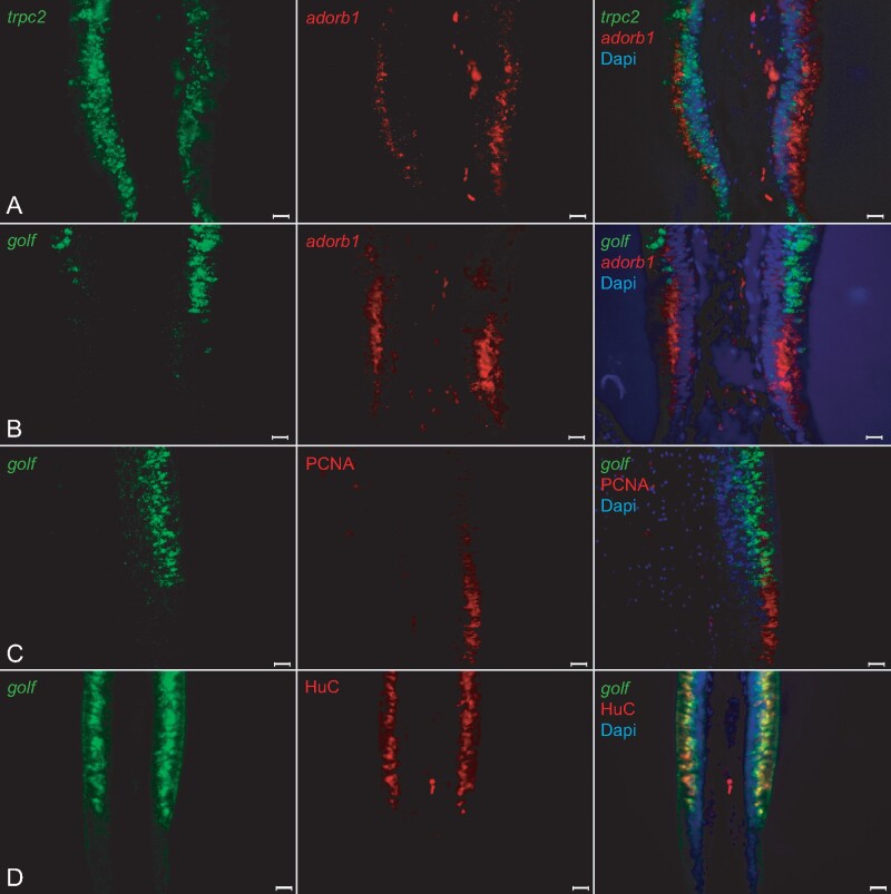
Fig. 7
Adorb1-expressing cells in lamprey are negative for several neuronal markers. Horizontal cross-sections of L. fluviatilis olfactory epithelium were double labeled with OSN markers in several combinations; nuclei are visualized with DAPI. Left and middle columns, single label; right column, merged pictures. Orientation of panels: the adorb1-positive region is always below the adjacent sensory region. All scale bars are 20 Ám. (A, B) Double ISH with neuronal markers for microvillous and ciliated OSNs (trpc2, and golf, respectively) and adorb1 shows no coexpression with either marker. (A) Unexpectedly, trpc2 (green) is expressed both in the sensory region and the adorb1-positive (red) segment. However, no double-labeled cells are seen. The nature of the trpc2-positive cells in the nonsensory region is unclear. (B) golf (green) is expressed in the sensory area, and exclusive of adorb1 expression (red). (C, D) Double labeling of golf with HuC and PCNA antibodies shows extensive overlap of golf and HuC at the cellular level, and complete absence of overlap for golf and PCNA, as expected.

