Figure 2
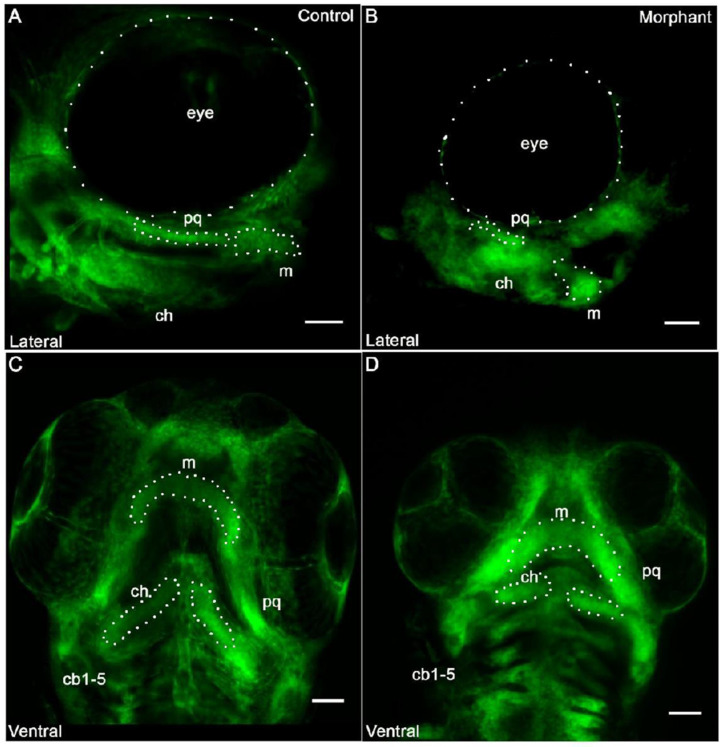
Cranial neural crest derived cells form segmented pharyngeal arches with abnormal shape under knockdown conditions of col11a1a. Fli1a:EGFP positive cells indicated that the neural crest derived cells were present within the pharyngeal arches. After occupying the pharyngeal arch, however, cells failed to form proper morphology. While the cells aligned to form an elongated structure in the palatoquadrate (pq) that joins the Meckel?s cartilage in the control morphant (A), the pq of the col11a1a morphant did not form the extended structure and the Meckel?s cartilage did not extend distally (B). The ventral view indicated segmentation of each of the developing pharyngeal arches in both the control (C) and the morphant (D). However, altered morphology was observed in the morphant (D). Scale bar 50 ?m. (m; Meckel?s cartilage; pq; palatoquadrate; ch; ceratohyal; cb; ceratobranchial).

