Figure 3
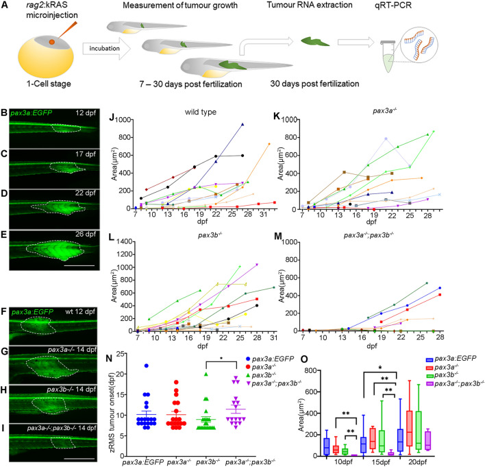
Functional analysis of zRMS progression in wild type and pax3 mutant zebrafish: (A) schematic illustration of generating zRMS, showing microinjection of rag2:kRASG12D into one cell stage zebrafish embryo, zRMS detection and monitoring of tumour growth from 7 to 30 dpf, tumour tissue excision and total RNA extraction at 30 dpf for gene expression analysis using qRT-PCR. (B?E) zRMS identified in pax3a:EGFP zebrafish transgenic line at 12 dpf, 17 dpf, 22 dpf, and 27 dpf. High intensity of GFP expression is observed at the tail area of the zebrafish larvae. Representative image of zRMS tumour progression (F) wild type at 12 dpf, (G) pax3a?/? at 14 dpf, (H) pax3b?/? at 14 dpf and (I) pax3a?/?;pax3b?/? at 14 dpf. Tumour area (?m2) of individual zRMS (J) wild type fish (n = 24), (K) pax3a?/? (n = 18), (L) pax3b?/? (n = 22), (M) pax3a?/?;pax3b?/? (n = 12). (N) zRMS tumour onset is indicated in days post fertilization in wild type fish (n = 20), pax3a?/? (n = 18), pax3b?/? (n = 22) and pax3a?/?;pax3b?/? (n = 16) zebrafish lines. (O) Tumour area (?m2) in zebrafish larvae that were injected with rag2-KRASG12D in wild type (n = 12), pax3a?/? (n = 10), pax3b?/? (n = 16) and pax3a?/?;pax3b?/? (n = 6) zebrafish lines at 10 dpf, 15 dpf and 20 dpf. All experiments were conducted in pax3a:EGFP background. Tumour areas are indicated with dashed lines. Error bars represent mean ± SEM and significance was calculated using Student t-test and Mann Whitney U test where p < 0.05 was considered significant, *p < 0.05, **p < 0.01, ***p < 0.001. Scale bar: 1 mm.

