
|
Fig. 6
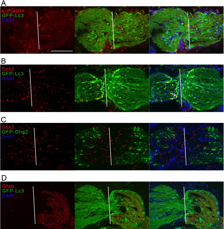
Fig. 6. Immunostaining for L-plastin, Sox2 and Gfap on optic nerve cryosections of Tg(CMV:GFP-Lc3) autophagy reporter fish and for Sox2 on optic nerve sections of Tg(olig2-GFP) fish, all tissues harvested at three days after optic nerve injury. (A) No overlap between the pan-leukocyte marker L-plastin (red) and Lc3 (green) could be detected in the injured optic nerve. (B) Lc3+ cells (green) overlapped with the Sox2 pluripotency marker (red), which could be expressed by glial precursor cells including OPCs, activated astroglia or newly differentiated oligodendrocytes during remyelination. See one double-positive cell indicated with an arrow. (C) OPCs and oligodendrocytes, positive for olig2 (green), did not show co-labeling with Sox2 (red). (D) Gfap (red), used to label astroglia and radial glia, and here possibly glial precursor cells, was strongly upregulated behind the site of impact at 3 dpi and overlapped partially with the autophagy reporter (green). Scale bar = 200 �m. Representative images of n = 4. The white line indicates the crush site. Dpi, days post-injury; Gfap, glial fibrillary acid protein; GFP, green fluorescent protein; Lc3, microtubule-associated protein 1A/1B-light chain 3; ONC, optic nerve crush; Sox2, sex determining region Y-box2. (For interpretation of the references to colour in this figure legend, the reader is referred to the web version of this article.)