
|
Fig. 1
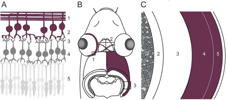
Fig. 1. Schematic representation of the studied components of the zebrafish visual system. (A) The first part of the visual system is the retina, in which the visual information flows from the photoreceptors (5) to the inner neurons (4) and eventually to the RGCs (2). The dendrites of the RGCs are located within the IPL (2), while the axons, in contrast, bundle inside the NFL (1) and form the optic projection. (B) From the retina (1), the optic projection, containing the optic nerve, optic chiasm and optic tract (all 2), innervates the optic tectum (3). (C) The two main domains of the zebrafish optic tectum are the PGZ (1) in which most of the cell bodies of the optic tectum are located, and the synaptic neuropil area (2–5, being the SEC, SGC, SFGS and SO, respectively). RGC axons mainly innervate the SO and SFGS (4–5). INL, inner nuclear layer; IPL, inner plexiform layer; NFL, nerve fiber layer; PGZ, periventricular gray zone; PRL, photoreceptor layer; RGC, retinal ganglion cell; RGCL, retinal ganglion cell layer; SAC, stratum album centrale; SFGS, stratum fibrosum et griseum superficiale; stratum opticum; SGC, stratum griseum centrale; SO, stratum opticum.