Image
Figure Caption
Fig. 1
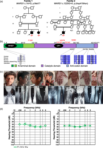
Pedigree, variant segregation, alignment of WARS1 orthologues with respect to protein domain structures, and clinical information. (a) Pedigrees of Families 1 (left) and 2 showing (right) segregation of the respective variant listed above each pedigree. Mutant alleles are represented with ??? and the reference allele is shown with ?+.? (b) Alignment of multiple WARS1 orthologues with amino acid substitutions involved in autosomal recessive (shown below the protein domain schematic) and autosomal dominant (shown above in red) illustrating amino acid conservation of the involved amino acid (boxed in black) and flanking region (autosomal recessively-associated alleles only). The domain diagram of human WARS1 shows the domain structures. CP1, connective polypeptide; WHEP, helix-turn-helix motif. (c) Facial features of the two affected individuals in Family 1 (IV:3 and IV:4) showing triangular face with micrognathia, and abundant hair and eyebrows, and large earlobes with apparent bifid tragus. Hands are small with round fingernails and show clinodactyly of the fifth finger, variation of deep palmar flexion creases, and presence of hypothenar creases. Radiology studies show mild medullary widening and minimal medial bowing of the mid/distal fibula shafts. Most severe, shown in IV:4, is coxa valga with deformity of the hips. (d) Pure-tone audiograms from the proband in Family 1 at the age of 19 years show mild, bilateral, sensorineural hearing loss. Right air conduction (circles), unmasked bone conduction (<), left air conduction (crosses) and unmasked bone conduction (>) are shown.
Acknowledgments
This image is the copyrighted work of the attributed author or publisher, and
ZFIN has permission only to display this image to its users.
Additional permissions should be obtained from the applicable author or publisher of the image.
Full text @ Hum. Mutat.

