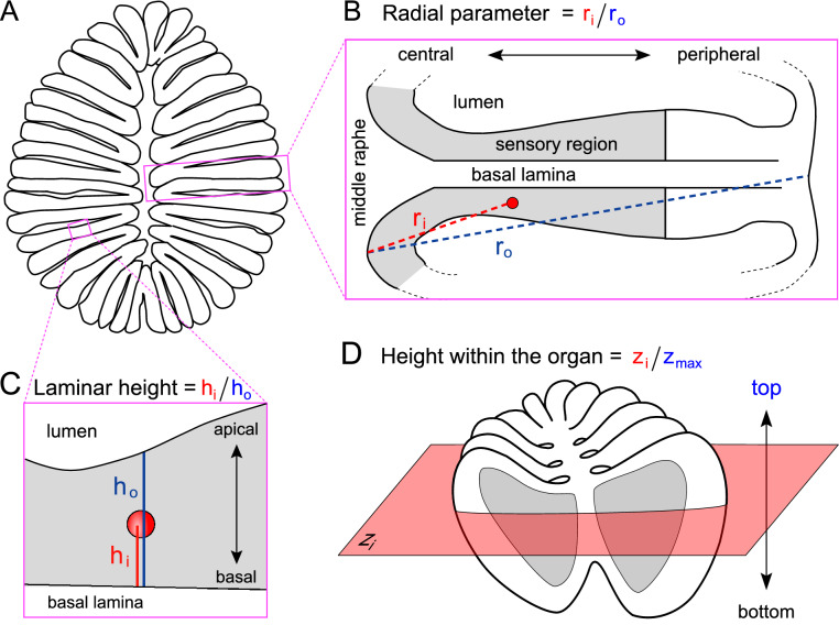
Fig. 2
Schematic representation of spatial coordinates measured. (A) Horizontal cross section of an olfactory epithelium. (B) Enlargement shows the radial parameter (ri), measured from middle raphe between lamellae to center of labeled cell and normalized to maximal radius (ro, including non-sensory area). One lamella is shown. (C) Enlargement shows the laminar height parameter (hi), measured from the border between basal lamina and the sensory layer to center of labeled cell and normalized to maximal height (ho, measured at position of labeled cell, as laminar height varies throughout the lamella). (D) The complete olfactory organ, all three spatial coordinates are shown. height-within-the-organ is measured as section number z, beginning with the most basal section still containing sensory surface (bottom to top).

