Fig. 7
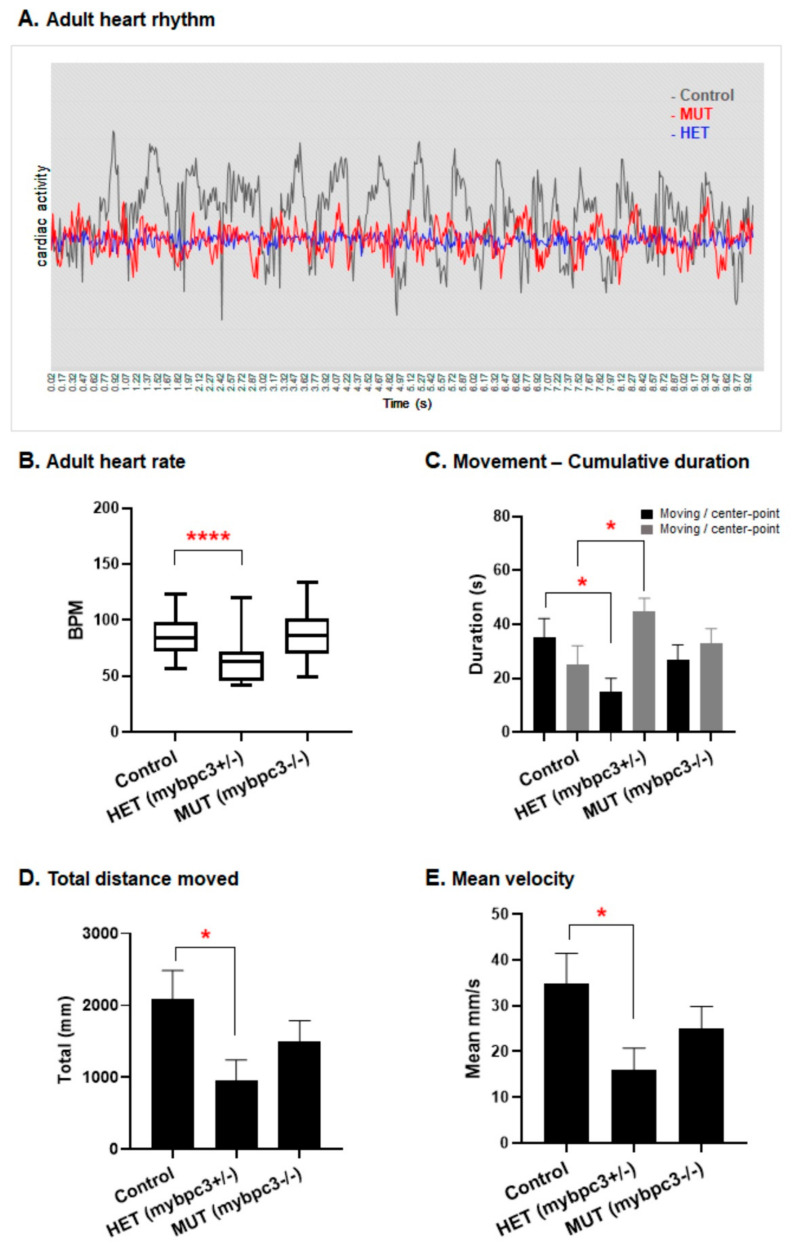
The zebrafish mybpc3 mutant adult heart function. (A) Cardiac activity percentage analyzed from oscillations in the cardiac signal at the contraction-relaxation status of the ventricle, with corresponding peaks and lows at regular intervals of contractions at the correct pace over time of a total of 10 s of video recording of beating heart. Both mybpc3+/? and mybpc3+/? groups displayed decreased amplitude. (B) Heart rate was measured in the adult zebrafish after incubation in the combined MS-222 (Tricaine methanesulfonate) and isoflurane for minimal cardiac consequence function effects. The examined adult heterozygous mybpc3+/? group (n = 35) displayed a significant reduction in heart rate, 58.36 beats per minute (BPM), **** p < 0.0001 in comparison to the control (n = 37) at 87.61 BPM and mutant mybpc3?/? (n = 40) at 88.51 BPM. The fluorescent heart of the fish was directly imaged for 10 s using a ZEISS stereomicroscope Lumar12 supplied with an Imaging source camera (Model33UX252); data are presented as mean ± SEM. (C) The calculated cumulative duration of moving rate to nonmoving was lower in the mybpc3 mutant fish. Similarly, the heterozygous mybpc3+/? group showed the lowest ratio decrease compared to the homozygous and control groups. (D,E) The maximum swimming speed and endurance experiments: the adult zebrafish mybpc3 mutant demonstrated declines in the exercise capacity. The total distance and velocity decreased significantly in the mybpc3 mutant fish. However, the heterozygous mybpc3+/? group showed a more significant decrease when compared to the control group. * p < 0.05.

