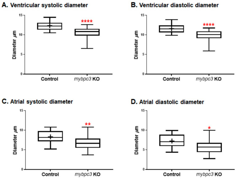
Fig. 3
The zebrafish mybpc3 knockout (KO) model displayed restrictive physiology of the cardiac chambers at 72 h post-fertilization (hpf). (A,B) Cardiac ventricular and (C,D) cardiac atrial analysis of the zebrafish mybpc3 KO demonstrated by the measurement of diastolic/systolic diameters that were significantly decreased in comparison to the wild-type (control) group. The number of embryos analyzed for the control = 28 and mybpc3 KO = 18. The larvae were mounted to image both chambers of the heart. Cardiac function analyses were represented in Box?Whisker plots and analysis using a one-way ANOVA multiple comparisons test. Values were expressed as the means ± SE. p values of <0.05 were considered statistically significant, a value of * p < 0.05, ** p < 0.01, and **** p < 0.0001.

