
|
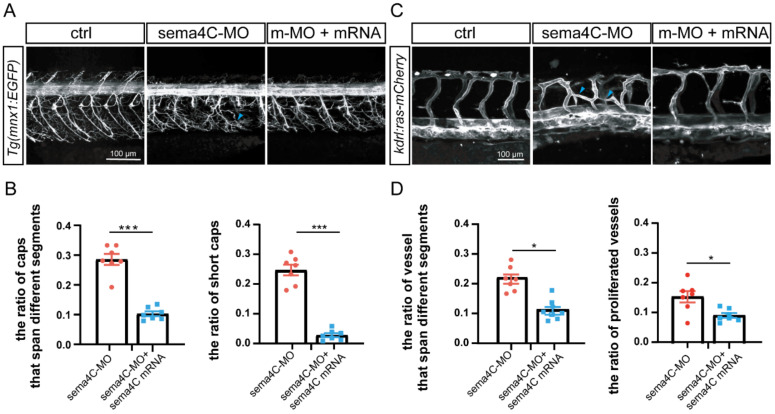
Fig. 6
Overexpressing sema4C partially rescued the abnormal of PMNs and ISVs in sema4C deficient embryos. (A). Confocal imaging analysis of PMNs in control and sema4C morphants at 72 hpf, blue arrowheads indicate aberrant PMNs and ISVs; (B). The statistical analysis of the proportion of deficient axonal trajectories of Caps (*** p = 0.0006) and short Caps (*** p = 0.0006) in the sema4C morphants and sema4C rescue groups at 72 hpf. Dots and squares represent the data of the experimental group and the rescue group, respectively. Mann?Whitney test; (C). Confocal imaging analysis of ISVs in control and sema4C morphants at 48 hpf; (D). The statistical analysis of the ratio of vessels that span different segments (* p = 0.035) and the proportion of proliferated vessels (* p = 0.0262) in the sema4C morphants and sema4C rescue groups at 48 hpf. Dots and squares represent the data of the experimental group and the rescue group, respectively. Mann?Whitney test.